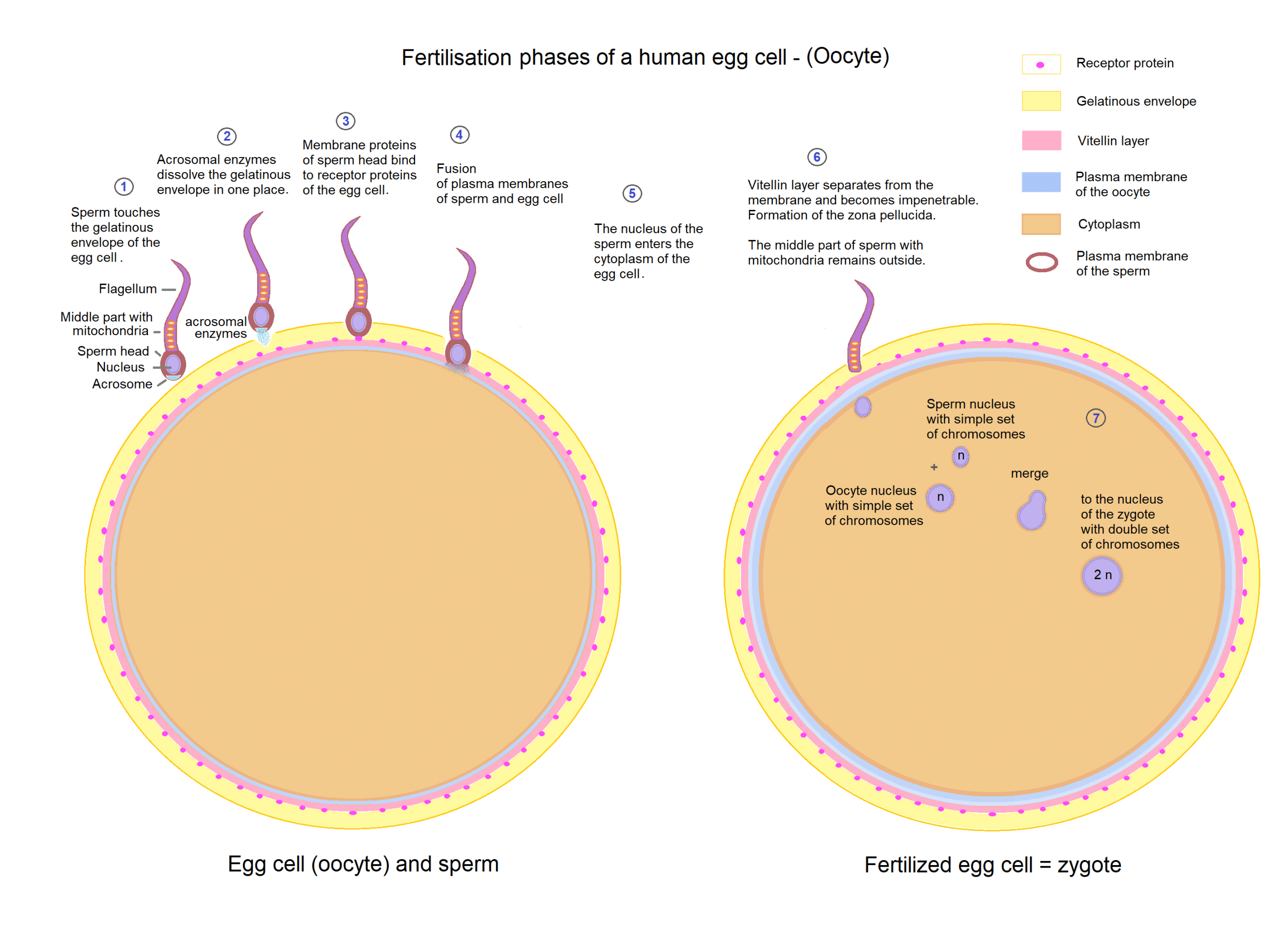
Conception, or fertilisation, describes the union of the male sperm and the female secondary oocyte (arrested in metaphase II) to form a zygote. The zygote subsequently divides within the fallopian tube and implants in the uterine wall leading to the establishment of pregnancy.
There are six important steps which must occur for fertilisation to be successful: sperm transport to the site of fertilisation, sperm capacitation, the acrosomal reaction, polyspermy block, completion of meiosis II and zygote formation.
In this article, we will go through the process of conception and highlight key learning points
Transport of Sperm
The passage of sperm through the female reproductive tract is highly regulated to maximise the chance of fertilisation by a sperm with normal morphology and vigorous motility.
After ejaculation, the semen immediately coagulates to form a loose gel. This serves to hold the sperm at the cervical os and to protect against the acidic pH and immunological response of the vagina. The gel is enzymatically degraded within one hour of deposition.
At the cervix, the sperm encounters cervical mucus. High levels of oestrogen around the time of ovulation reduce its viscosity and alter its microstructure to allow for easier passage of the sperm through the cervix and into the uterus. Additionally, components of the seminal fluid assist the sperm to penetrate the mucus barrier. Cervical mucus presents a greater barrier to sub-motile sperm and is thus a means of sperm selection.
Once the sperm enters the uterus, pro-ovarian contractions of the uterine myometrium help to propel the sperm towards the fallopian tube. Prostaglandins present in the seminal fluid promote these myometrial contractions. Once in the fallopian tube, the sperm is no longer at risk of attack by immune cells of the vagina, cervix and uterus.
The first sperm enter the fallopian tubes only minutes after ejaculation, however, may survive in the female reproductive tract for up to five days.
Sperm Capacitation
For fertilisation to occur, the sperm must undergo a process called capacitation. Capacitation is the final maturation process of the sperm, whereby physiological changes to the sperm render them capable of fertilisation.
Capacitation is triggered by uterine secretions which act to destabilise the plasma membrane of the sperm. Specific structural changes include:
- Cholesterol efflux – cholesterol acceptors in the extracellular matrix remove cholesterol from the plasma membrane of the sperm. This changes the cholesterol-phospholipid ratio and thus alters membrane fluidity.
- Redistribution of glycoproteins and change/activation of proteins on the plasma membrane of sperm head
- Increase in the number of CatSper Channels (cation channel of the sperm) on the flagellum
Sperm capacitation has two important outcomes. Firstly, it prepares the sperm’s head for the acrosomal reaction. Secondly, the motility of the sperm is hyperactivated by the changes to the strength and speed of the flagellum’s movements, induced by the increase in CatSper channels.
CatSper channels allow for Ca2+ influx, with downstream activation of cAMP and PKA. This gives the sperm the additional strength required to reach the secondary oocyte.
Sperm-Oocyte Interactions
Once the capacitated sperm reach the secondary oocyte in the fallopian tube, multiple sperm-oocyte interactions must occur before the sperm can fertilise the egg.
The Cumulus-Oocyte Complex
The cumulus-oocyte complex (COC) describes an oocyte surrounded by specialised granulosa cells known as cumulus cells. The capacitated sperm first encounters the cumulus-oocyte complex surrounding the secondary oocyte and a much higher concentration of follicular fluid.
Hyaluronidase present on the surface of the capacitated sperm head dissolves the hyaluronic acid, cementing the cells of the COC together, and enabling the capacitated sperm to penetrate the COC.
The Zona Pellucida
The sperm then reach a layer of glycoproteins surrounding the oocyte, known as the zona pellucida (ZP). There are four main cell surface glycoproteins making up the zona pellucida (ZP1, ZP2, ZP3, ZP4) – ZP3 proteins will only bind with receptors on the plasma membrane of the capacitated human sperm.
This lock-and-key type mechanism is species-specific and prevents the sperm and egg of different species from fusing.
The Acrosomal Reaction
Binding to ZP3 causes the sperm’s plasma membrane to fuse with its acrosomal membrane, leading to the release of hydrolytic and proteolytic enzymes from the sperm’s acrosome. These enzymes are responsible for digesting the zona pellucida, enabling the sperm to reach the cell membrane.
Multiple sperm are involved in the above three processes. However, only one of these sperm will fuse and fertilise the oocyte.
Sperm-Oocyte Fusion
The Cortical Reaction
After the ZP is digested, a single sperm fuses with the cell membrane of the oocyte. This causes the smooth endoplasmic reticulum of the oocyte to release Ca2+, raising intracellular Ca2+ levels. This is called the cortical reaction.
There are three important outcomes of the cortical reaction:
- Slow polyspermy block
- Completion of meiosis II
- Zygote formation.
We will now explore these in more detail.
Polyspermy Block
To prevent multiple sperm from binding to the cell membrane, there are two mechanisms of polyspermy block.
Fast Block: Na+ dependent, rapid onset, short duration
The first mechanism of polyspermy block is the fast block. Binding of sperm to the cell membrane activates Na+ ion channels which allow for Na+ influx into the egg. This causes depolarisation, making the oocyte more positive and thus repelling further sperm and inhibiting them from binding. A fast block has a rapid onset, but short duration.
Slow Block: Ca2+ dependent (occurs secondary to the cortical reaction), slow onset, long duration
The second mechanism of polyspermy block is called the slow block, which occurs as part of the cortical reaction. Fusion of the sperm and oocyte cell membrane activates the smooth endoplasmic reticulum (smooth ER), causing it to release large amounts of Ca2+. This stimulates intracellular lysosomes to release hydrolytic enzymes to further degrade the ZP, and stimulates exocytosis of cortical granules to harden the oocyte cell membrane, making it impenetrable to further sperm.
The slow block has a slower onset but is longer in duration.
Completion of Meiosis II
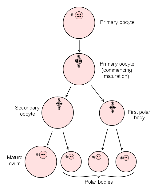
The Ca2+ released during the cortical reaction acts at the nucleus of the secondary oocyte to stimulate the completion of meiosis II. As a result, there is a definitive ovum and a polar body; the latter is later degraded.

Fig 2 – Oogenesis
Zygote Formation and Implantation
Finally, the pronucleus of the definitive ovum (n) is now able to fuse with the pronucleus of the sperm cell (n). This produces the zygote (2n).
The zygote continues to divide via mitosis within the fallopian tubes. At the 16-cell stage (3-4 days after fertilisation), the embryo is called a morula. All 16 cells of the morula are totipotent, and not specified to a particular fate.
At day 5, the embryo becomes a blastocyst, characterised by the development of a distinct inner cell mass and blastocoele (fluid-filled cavity), and surrounded by trophoblast cells. Implantation occurs on days 7-8, at the late blastocyst stage.
For more information on the early stages of embryonic development, see here.

