A reflex is defined as an involuntary, unlearned, repeatable, automatic reaction to a specific stimulus which does not require input from the brain. The muscle stretch reflex is the most basic reflex pathway in the body and as such, understanding this allows understanding of more complex reflexes.
This article shall discuss the components of a reflex arc, the monosynaptic reflex and relevant clinical issues.
Reflex Arc Components
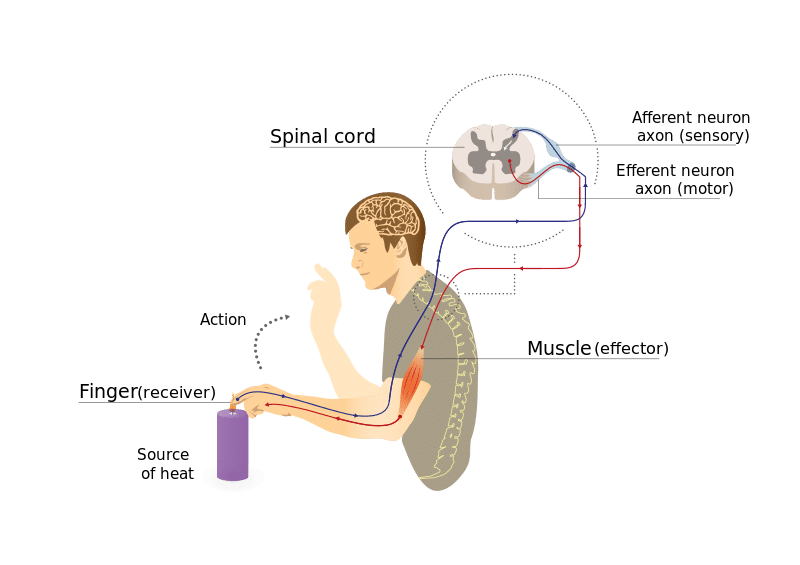
A reflex arc is a neural pathway that controls a reflex. Most sensory neurones have a synapse within the spinal cord. This allows for reflexes to take place without the involvement of the central nervous system (CNS) – speeding up the process. The pathway can be described as a ‘reflex arc’ which is made up of 5 components:
- A receptor – muscle spindle
- An afferent fibre – muscle spindle afferent
- An integration centre – lamina IX of spinal cord
- An efferent fibre – α-motoneurones
- An effector – muscle
The Monosynaptic Stretch Reflex
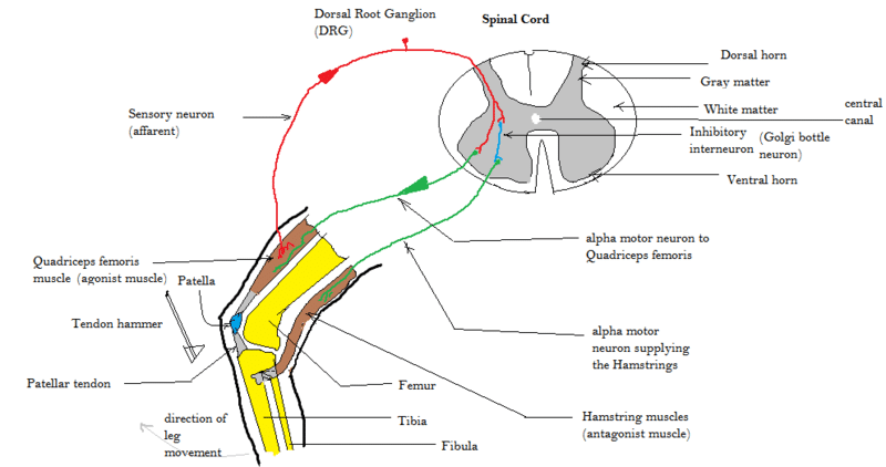
A monosynaptic reflex, such as the knee jerk reflex, is a simple reflex involving only one synapse between the sensory and motor neurone.
The pathway starts when the muscle spindle is stretched (caused by the tap stimulus in the knee jerk reflex). The muscle spindles are responsible for detecting the length of the muscles fibres.
When a stretch is detected it causes action potentials to be fired by Ia afferent fibres. These then synapse within the spinal cord with α-motoneurones which innervate extrafusal fibres. The antagonistic muscle is inhibited and the agonist muscle contracts i.e. in the knee jerk reflex the quadriceps contract and the hamstrings relax.
The sensitivity of the reflex is regulated by gamma motoneurones. These lead to tightening or relaxing of muscle fibres within the muscle spindle. It is thought that this takes place to allow preservation of the stretch reflex when muscles are contracted, although not much is known about it.

Fig 2 – Diagram showing the muscle stretch reflex using the knee jerk reflex as an example.
Clinical Relevance – Testing Reflexes
When testing reflexes it is important to know which spinal root level you are testing.
If the reflex is not present it could be due to a problem with the receptor, the spinal cord, the motoneurone, the neuromuscular junction or the muscles. It is also important to look for hyperreflexia (UMN sign) or hyporreflexia (LMN sign)
| Reflex | Spinal Levels |
| Biceps reflex | C5/C6 |
| Brachioradialis reflex | C6 |
| Extensor digitorum reflex | C6/C7 |
| Triceps reflex | C6-C8 |
| Patellar reflex | L2-L4 |
| Achilles reflex | S1/S2 |
The strength of the reflex can be graded from 0 (no response) to 4+ (clonus). 2+ (brisk response) is normal.

