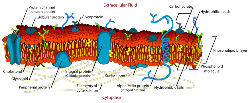
Cell membranes are an essential component of the cell, providing separation between the intracellular and extracellular environment. They are composed of lipids, proteins and carbohydrates.
In this article, we shall consider the main functions of the cell membrane, the composition of membranes and clinical conditions in which a portion of the cell membrane is abnormal.
Structure of the Cell Membrane
A simplified rough guide of the dry weight is shown in Table 1.
| Dry Weight |
| 40% lipid
– E.g. phospholipid molecules and cholesterol |
| 60% protein
– E.g. channel proteins and carrier proteins |
| 1-10% carbohydrate
– Often found attached to proteins/lipids on the outside of the cell membrane – a coat of carbohydrate surrounding a cell is often called the glycocalyx |
Phospholipids
The membrane bilayer contains many kinds of phospholipid molecules, with different-sized head and tail molecules.
These consist of a head molecule, a phosphate molecule, a glycerol and two fatty acid chains.
- Head group – This is a polar group e.g. a sugar or choline, meaning that the head end of the phospholipid is hydrophilic.
- Tail of 2 fatty acid chains – normally consisting of between 14-24 carbons (but the most common carbon lengths are 16 and 18). If the chain contains a cis double bond then the chain is kinked – therefore reducing the tight packing of the membrane and so increasing its movement. As the tail is made of fatty acids, it does not form hydrogen bonds with water and therefore is hydrophobic and non-polar.
Phospholipid molecules are therefore amphipathic – being both hydrophilic and hydrophobic. They spontaneously form bilayers in the water with the head groups facing out and the tail groups facing in.
In the bilayer, there are van der Waal forces between the fatty acid tails of the phospholipid, with electrostatic and hydrogen bonds between the hydrophilic groups and water.
Cholesterol
Cholesterol is vital for many functions in a cell, including very importantly, a major constituent of the cell membrane.
Cholesterol itself consists of a polar head, a planar steroid ring and a non-polar hydrocarbon tail. Cholesterol is important in the membrane as it helps to maintain cell membrane stability and fluidity at varying temperatures.
Cholesterol is bound to neighbouring phospholipid molecules via hydrogen bonds and therefore at low temperatures, reduces their packing. Overall this means at low temperatures, when the rate of movement is lowest, a fluid phase is maintained.
At high temperatures, cholesterol helps to stop the formation of crystalline structures and the rigid planar steroid ring prevents intrachain vibration and therefore making the membrane less fluid.
Membrane Proteins
As was shown in the table above, a typical cell membrane consists of around 60% protein. There is such a high proportion of protein because they are so vital in almost every process within a cell. A list of just a few functions of membrane proteins could include:
- Catalysts – enzymes.
- Transporters, pumps and ion channels.
- Receptors for hormones, local mediators and neurotransmitters.
- Energy transducers.
More active cells or organelles e.g. mitochondria, tend to contain more proteins, showing again that the function determines structure.
As part of the cell membrane, proteins can either be deeply embedded within the bilayer (integral) or be associated with the surface of the cell (peripheral).
Functions of the Cell Membrane
Cell membranes are vital for the normal functioning of all the cells in our bodies. Their main functions consist of:
- Forming a continuous, highly selectively permeable barrier – both around cells and intracellular compartments.
- Allowing the control of an enclosed chemical environment – important to maintain ion gradients.
- Communication – both with the extracellular and extra-organelle space.
- Recognition – including recognition of signalling molecules, adhesion proteins and other host cells (very important in the immune system).
- Signal generation – in response to a stimulus creating a change in membrane potential.
In a cell, different parts of the membrane have different functions and therefore their structure is specialised for this. An example of this specialisation can be seen in the different parts of a nerve; the cell membrane in the axon is specialised for electrical conduction whereas the end of the nerve is specialised for synapsing, meaning the composition of the membrane is different.
Clinical Relevance – Hereditary Spherocytosis
Hereditary spherocytosis is a condition in which spectrin, a peripheral cytoskeletal protein, is depleted by 40-80%. There are both autosomal dominant and recessive forms of the condition, with differing severity.
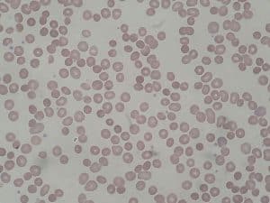
As a result of this lack of spectrin, erythrocytes cannot effectively maintain their biconcave structure, and assume a spherical shape. This decreases their ability to travel through the microvasculature of the body and results in increased erythrocyte lysis.
There are 3 other types of spherocytosis that result from defects in ankyrin, band 3 and protein 4.2, however, spectrin is the most significant.
The signs and symptoms of the condition include:
- Mild to moderate anaemia
- Possible jaundice
- Possible splenomegaly


