Neurones communicate with each other via electrical signals known as action potentials. They are brief changes in the voltage across the membrane due to the flow of certain ions into and out of the neurone. In this article, we will discuss how an action potential (AP) is generated and how its conduction occurs.
The Resting Membrane Potential
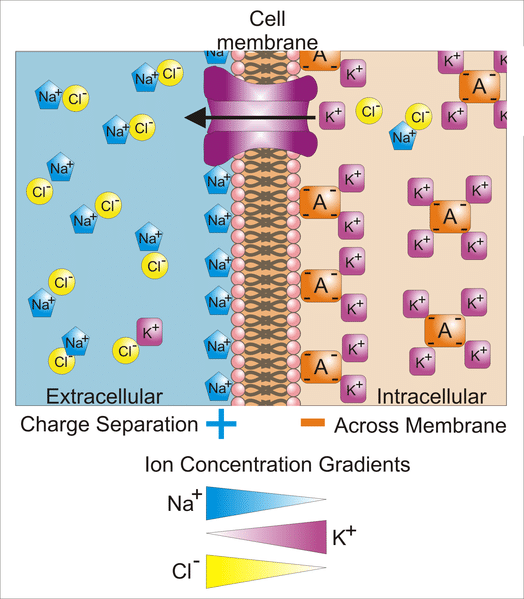
The resting membrane potential of cells varies depending on the cell type. For neurones, this typically sits between -50 and -75mV. This value depends on the types of ion channels that are open and the concentrations of different ions in the intracellular and extracellular fluids during the resting state. In neurones, K+ and organic anions are typically found at a higher concentration within the cell than outside, whereas Na+ and Cl- are typically found in higher concentrations outside the cell.
This difference in concentrations provides a concentration gradient for ions to flow down when their respective channels are open. Hence, K+ ions would be moving out of the cells, while Na+ and Cl- ions would be moving into the cell. At the resting state, the cell is mostly permeable to K+, as such this exerts the greatest influence on the resting membrane potential out of the three ions. Further information on the resting potential generation can be found here.
These concentration gradients are maintained by the action of the Na+/K+ ATPase via active transport, which in turn maintains the membrane potential.
Generation of Action Potentials
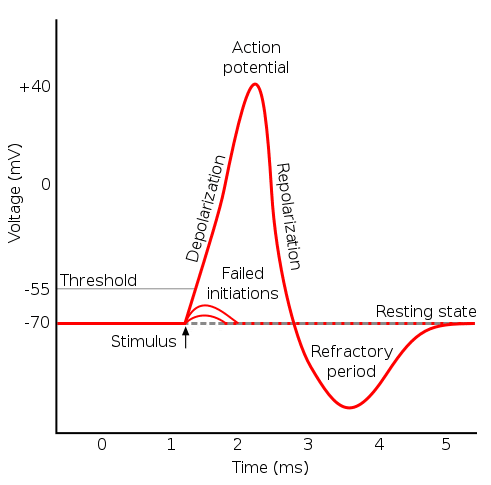
During the resting state, the membrane potential arises because the membrane is predominantly permeable to K+. An action potential begins at the axon hillock as a result of depolarisation. During depolarisation, voltage-gated sodium ion channels open due to an electrical stimulus. As the sodium ions rush back into the cell, their positive charge changes potential inside the cell from negative to more positive.
If a threshold potential is reached, then an action potential is produced. Action potentials will only occur if a threshold is reached. Additionally, if the threshold is reached, then the response of the same magnitude is always elicited, irrespective of the strength of the stimulus. Hence, action potentials can be described as “all-or-nothing“.
Once the cell has been depolarised the voltage-gated sodium ion channels begin to close. The positive potential inside the cell causes voltage-gated potassium channels to open and K+ ions now move down their electrochemical gradient out of the cell. As the K+ moves out of the cell, the membrane potential becomes more negative and starts to approach the resting potential.
Typically, repolarisation overshoots the resting membrane potential, making the membrane potential more negative. This is known as hyperpolarisation. It is important to note that the Na+/K+ ATPase is not involved in the repolarisation process following an action potential.
The Refractory Period
Every action potential is followed by a refractory period. This period can be further divided into:
- The Absolute Refractory Period which occurs once the sodium channels close after an AP. Sodium channels then enter an inactive state during which they cannot be reopened, regardless of the membrane potential.
- The Relative Refractory Period which occurs when sodium channels slowly come out of the inactivation. During this period the neurone can be excited with stimuli stronger than the one normally needed to initiate an AP. Early on in the relative refractory period, the strength of the stimulus required is very high. Gradually, it becomes smaller throughout the relative refractory period as more sodium channels recover from the inactivation stage.
Propagation of Action Potentials
Action potentials are propagated along the axons of neurones via local currents. Local currents induce depolarisation of the adjacent axonal membrane. Where this reaches a threshold, further action potentials are generated. The areas of the membrane that have recently depolarised will not depolarise again due to the refractory period – meaning that the action potential will only travel in one direction.
These local currents would eventually decrease in charge until a threshold is no longer reached. The distance that this would take depends on the membrane capacitance and resistance:
- Membrane Capacitance – The ability to store charge. The lower capacitance results in a greater distance before the threshold is no longer reached.
- Membrane Resistance – This depends on the number of ion channels open. The lower the number of channels open, the greater membrane resistance is. A higher membrane resistance results in a greater distance before the threshold is no longer reached.
Myelinated Axons
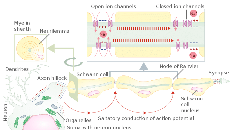
In order to allow rapid conduction of electrical signals through a neurone and make them energy-efficient, certain neuronal axons are covered by a myelin sheath. The myelin sheath surrounds the axon to form an insulating layer. Further information on the myelin sheath can be found here.
Along a myelinated axon, there are periodic gaps where there is no myelin and the axonal membrane is exposed. These gaps are called Nodes of Ranvier. In contrast to myelinated sections of the axon that lack voltage-gated ion channels, nodes of Ranvier harbour a high density of ion channels. This means that an action potential can only occur at the nodes.
The myelin sheath speeds up the conduction by increasing the membrane resistance and reducing the membrane capacitance. Hence, the action potential is able to propagate along the neurone at a higher speed than would be possible in unmyelinated neurons. The electrical signals are rapidly conducted from one node to the next, where is causes depolarisation of the membrane. If the depolarisation exceeds the threshold, it initiates another action potential which is conducted to the next node. In this manner, an action potential is rapidly conducted down a neurone. This is known as saltatory conduction.

Fig 3 – Diagram to show how the myelin sheath results in saltatory conduction of an action potential along an axon.
Clinical Relevance – Multiple Sclerosis
Multiple sclerosis (MS) is an acquired, chronic autoimmune disorder affecting the CNS. It results in demyelination, gliosis, and neuronal damage. Common presentations of the disease are optic neuritis, transverse myelitis, and cerebellar symptoms such as ataxia.

Fig 4 – Diagram demonstrating the main symptoms that multiple sclerosis may present with.
There are three main patterns of disease:
- Relapsing-remitting – Patients face episodes of remission (during which no symptoms are present) and exacerbations of the disease.
- Secondary Progressive – Initially the MS is of a relapsing-remitting pattern. However, at some point, the disease course changes, and the neurological function gradually worsens.
- Primary progressive – After the onset of the disease there is a steady progression and worsening of the disease.
There is no known cure for MS. However some therapies have proven useful in terms of managing acute exacerbations, preventing exacerbations, and slowing down disability. For example, high doses of intravenous corticosteroids can help to relieve symptoms in acute exacerbations.


