- Biochemistry
- Histology
- Cardiovascular
- Respiratory
- Gastrointestinal
- Urinary
- Reproductive
- Neurology
- Endocrine
- Immunology/Haematology
Cardiac Cycle
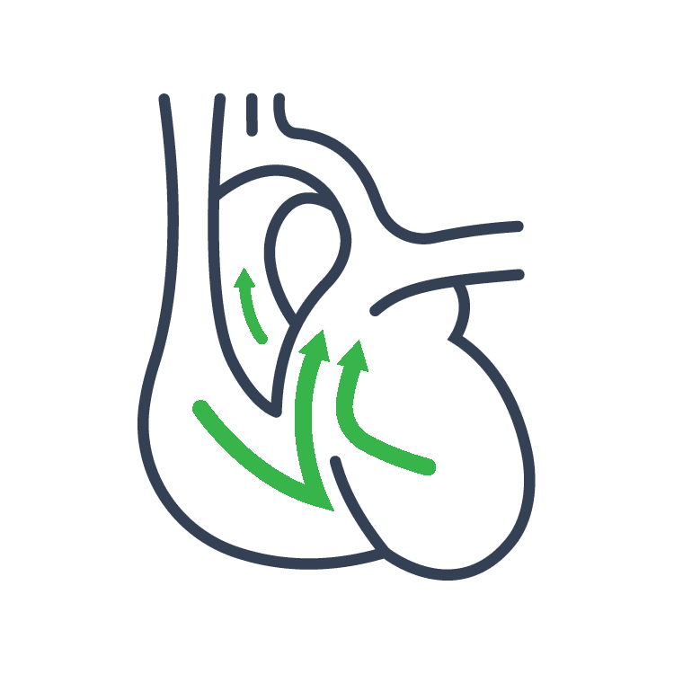
The first article in this section regards the cardiac cycle in overview. At rest the heart pumps around 5L of blood around the body every minute, but this can increase massively during exercise. In order to achieve this high output efficiently the heart works through a carefully controlled sequence with every heart beat – this sequence of events is known as the cardiac cycle. In this article, we will consider the changes that occur throughout each cycle to ensure that adequate perfusion is delivered to the tissues of our body.
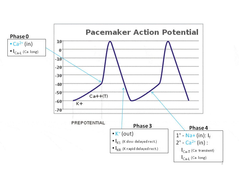
Our next article describes the physiology of the pacemaker cells of the cardiac tissue. In the heart, electrical impulses are generated by specialised pacemaker cells and spread across the myocardium in order to produce a coordinated contraction in systole. The action potential generated is a characteristic disturbance of the potential difference between the inside and the outside of the cell. The particular action potential generated by cardiac pacemaker cells is very different to that in nerve and striated muscle cells, and to that of ventricular myocardial cells. In this article we shall consider cardiac pacemaker cells and the action potential they produce in more detail.
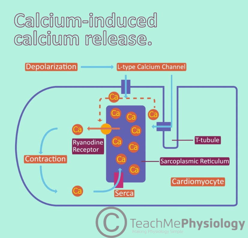
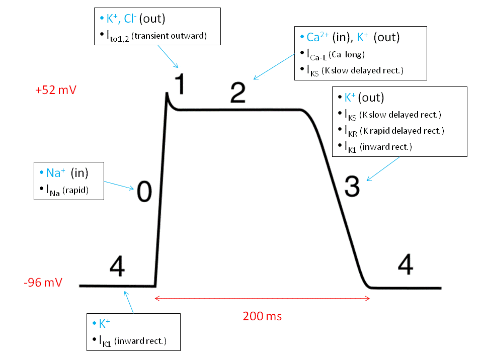
The final article in this section will consider the action potentials found within ventricular cells. Action potentials in ventricular myocytes trigger the Ca2+ entry that is necessary for their contraction. Their synchronicity, characteristic shape and length safeguards the heart against abnormal electrical activity. When these safeguards go wrong it can be potentially life threatening. In this article we will look at how action potentials spread in ventricular cells, their shape and modulation in disease states.


