Epithelial cells are the cellular components of the epithelium (pleural: epithelia). Epithelia are layers of contiguous cells that line the surfaces of organs and tissues.
In this article, we will consider the different types of epithelia, the different types of epithelial cell and discuss some clinical applications of this physiology.
Overview
Epithelial cells are localised to three distinct areas of the body. They can be found:
- Covering the whole external surface of the body, as part of the skin.
- Lining interior tracts which open to the exterior, such as the respiratory, gastrointestinal and genitourinary tracts.
- Lining interior enclosed spaces, such as blood vessels, the peritoneum, the pleura and the pericardial sac.
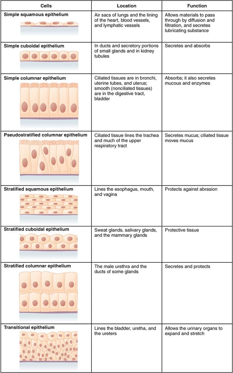
Epithelial cells are classified in two ways. Firstly they can be classified depending on how many layers of cells are present, and secondly, by shape.
Clinical Relevance – Cancer
While histology may sometimes seem irrelevant to clinical practice, epithelial cells types are important for classifying cancers. Knowing what sort of epithelium is present in certain organs can allow you to predict the most common cancer type affecting that organ.
For example, the primary form of oesophageal cancer is squamous cell carcinoma, reflecting the fact that the oesophagus is lined with stratified squamous epithelium.
Simple vs. Stratified
Simple epithelia are defined as epithelia which are one cell layer thick – i.e. every cell attaches to the basement membrane. The basement membrane is a thin but strong, acellular layer which lies between the epithelium and the adjacent connective tissue. It is sometimes referred to as the “basal lamina”.
The basement membrane is clinically important in cancer, as the degree to which malignant cells penetrate it is highly relevant to prognosis. The side of the cell furthest from the basement membrane is known as the apical border.
Stratified epithelia consist of multiple layers of cells, with one layer anchored to the basement membrane, known as the basal layer.
Cell Shape
Epithelial cells are also classified by their shape:
- Squamous – may be simple or stratified.
- Cuboidal – may be simple or stratified.
- Columnar – may be simple or stratified.
- Pseudostratified – always simple.
- Transitional (urinary) – always stratified.
Note that all epithelia, whether simple or stratified, are avascular, receiving oxygen via diffusion.
We will now look at some examples of these and how their structure relates to their function.
Simple Epithelia
Simple Squamous Epithelium
Simple squamous epithelium comprises a single layer of flattened cells, making it the thinnest sort of epithelium.
Fig 2 – Diagram of simple squamous epithelium
It is found in multiple locations, and provides various functions, most of which relate to its thinness.
| Location | Function | Notes |
| Alveoli of the lungs | Gas exchange | – |
| Bowman’s Capsule and Loop of Henle of kidney | Barrier for filtration | – |
| Lining of the blood and lymph vessels | Exchange of gases and nutrients.
Passage of certain blood cells into tissues. |
The simple squamous epithelia lining the blood and lymph vessels is known as “endothelium” |
| Lining of the body cavities – i.e. the pleura, pericardium and peritoneum | Lubrication between tissues and organs | The simple squamous epithelia lining the body cavities is known as “mesothelium” |
Simple Cuboidal Epithelium
Simple cuboidal epithelium comprises a single layer of cuboidal, or roughly square, cells.
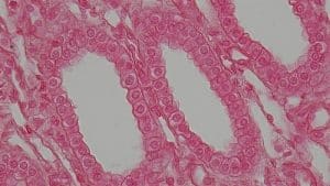
Fig 3 – Simple Cuboidal Epithelium
Again, it is found in various locations and has varying functions:
| Location | Function |
| Thyroid follicles | Hormone synthesis, storage and mobilisation |
| Small ducts of exocrine glands | Absorption and passage of exocrine secretions |
| Kidney tubules | Absorption and secretion |
| Surface of ovary (“germinal epithelium”) | Barrier/covering of follicles |
Simple Columnar Epithelium
Simple columnar epithelium comprises a single layer of long, thin cells.
Some simple columnar cells also have cilia (hair-like projections) or microvilli (finger-like projections) protruding from their apical borders, which are further specialisations that aid certain functions.
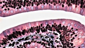
Fig 4 – Simple columnar epithelium
Examples of their location and functions include:
| Location | Function | Specialisations |
| Lining of the stomach and gastric glands | Absorption and secretion of gastric juices | Microvilli form a “brush border” which increases surface area for absorption |
| Small intestine and colon | Absorption, secretion and lubrication | Microvilli form a “brush border” which increases surface area for absorption |
| Gallbladder | Absorption of water and electrolytes from the bile | – |
| Fallopian tubes | Transport of ova | Some cells are ciliated to help to waft the egg along from the ovary to the uterus |
Pseudostratified Epithelium
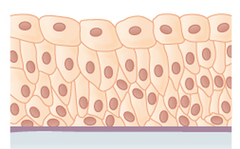
Pseudostratified epithelium is so-called because at first glance the cells appear to be several layers thick. However, every cell attaches to the basement membrane and therefore it is defined as simple epithelium.
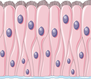
Fig 5 – Diagram of pseudostratified epithelium
Examples of its location and functions include:
| Location | Function | Specialisations |
| Lining of the nasal cavity, trachea and bronchi | Secretion of mucus, trapping of particles, and removal of mucus. | Cilia aid the passage of mucus |
| Epididymis and vas deferens | Absorption of fluid, secretion of substances which promote sperm maturation, and passage of sperm one motile. | Stereocilia (very long microvilli) aid the passage of sperm |
Stratified Epithelium
Stratified Squamous Epithelium
Stratified squamous epithelium can be further divided into keratinised and non-keratinised.
Keratinised stratified squamous epithelium is multi-layered squamous epithelium where the upper layers of cells, furthest from the basement membrane, are no longer alive and are filled with a protein called keratin.
Keratinised stratified squamous epithelium forms the epidermis of the skin, and a small amount is also found in the mouth. Its functions are to:
- Protect against physical trauma and abrasion
- Prevent water loss
- Provide a physical barrier against the invasion of microbes
- Protect against UV light
Non-keratinised stratified squamous epithelium is primarily involved in protecting against abrasion, and reducing water losses, thus keeping surfaces moist.
It is found in a wider variety of locations than keratinised stratified squamous epithelium, including the vagina, oesophagus, larynx, mouth, cornea, and part of the anal canal.
In the vagina, the epithelial cells are also involved in the maintenance of a low pH. The cells are rich in glycogen which acts as a substrate for lactobacilli to produce lactic acid, thus lowering pH.
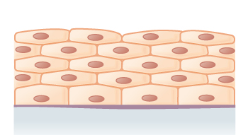
Fig 6 – Diagram of stratified squamous epithelium
Transitional Epithelium (aka “Urinary Epithelium”)
Transitional epithelium is found solely in the urinary tract. It changes shape in response to stretch. In its relaxed state, the cells usually appear more cuboidal or columnar, but as it stretches, they flatten out, appearing more like squamous cells.
Transitional epithelium is present in the renalcalyces, ureters, bladder and urethra. Its unique appearance allows distensibility, but it is also involved in protecting the underlying tissues from toxic chemicals found in the urine.

