The endoplasmic reticulum is the major site of synthesis in the cell. It is a system of flattened sacs (cisternae) that are continuous with the outer nuclear envelope. Its physiological function has a very close association with that of the Golgi apparatus and together, they form the secretory pathway of the cell.
The endoplasmic reticulum is classified as either rough or smooth, with minor variations in size and function in specialised tissue. In this article, we will look at the structure and function of the rough and smooth endoplasmic reticulum, and consider some clinical relevance.
Rough Endoplasmic Reticulum (RER)
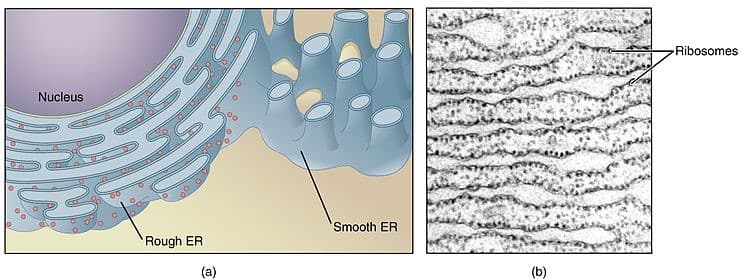
The rough endoplasmic reticulum (RER) takes its name from the many ribosomes attached to the cytoplasmic surface. The RER takes developing proteins from the cytosol and continues their development prior to completion in the golgi apparatus.
Proteins that move across the membrane of the RER undergo a series of post-translational modifications, including the addition of signal sequences to target them to the correct part of the cell. Cells that produce many secretory proteins will have will extensive RER and mitochondria.
The RER plays an important role in the synthesis of proteins that are destined for:
- Secretion into the extracellular matrix e.g. mucus and enzymes.
- Association with the cell membrane e.g. receptors and channels.
- Membrane-bound vesicles e.g. enzymes of lysosomes.
Part of the modification process involves the folding of developing proteins. Correctly folded proteins are transported to the golgi for secretion. Incorrectly folded proteins are kept within the cell and eventually destroyed.
Despite the presence of ribosomes being such a defining feature of RER, RER-ribosomal interactions are not permanent and thus go through periods of attachment and detachment depending on the demand for protein secretion. Adhesion to the surface is energy-dependent and so we see ribosomes detach in hypoxic cell injury when ATP/GTP synthesis is reduced.
Smooth Endoplasmic Reticulum (SER)
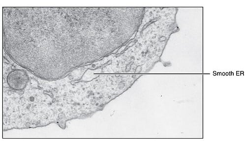
The smooth endoplasmic reticulum is important in the synthesis of lipids, phospholipids, and steroids. It is often less extensive and ribosomes do not associate with it, though certain specialised tissues (e.g. steroidogenic cells and muscles) often have extensive SER.
They also contain cytochrome P450 enzymes which are important in the metabolism of certain drugs and toxins e.g. alcohol and barbiturates. Hepatocytes store glycogen in regions that are rich in SER.
The smooth endoplasmic reticulum found within muscle is known as the sarcoplasmic reticulum and serves a specialised function.

Fig 2 – Electron microscopy of the Smooth Endoplasmic Reticulum
Sarcoplasmic Reticulum
The SER in muscle is extensive because it plays a major part in the sequestration of calcium, which modulates the tonic force of contraction (and relaxation).
The ultrastructure of the sarcoplasmic reticulum differs between muscle types. In striated muscle, they are arranged around the perpendicular T tubule, where the action potential/ surface Ca2+ inflow can initiate the calcium spike. This gives rise to the diads (cardiac muscle) and triads (skeletal muscle). Different types of muscles have different extensiveness of SR.
Further information on the role of the sarcoplasmic reticulum in muscle contraction can be found here.
Clinical Relevance – Malignant Hyperthermia
Malignant hyperthermia (MH) is an acute condition that arises as a result of a mutation of the ryanodine receptor of the sarcoplasmic reticulum in the muscle. The mutation leads to an increased sensitivity of the receptor to any potential agonist resulting in an exaggerated release of calcium resulting in what is known as a hypermetabolic crisis – a rapid acceleration of muscle metabolism with massive consumption of oxygen.
The factor causing fatality is the rapid rise of body temperature that accompanies accelerated oxidative phosphorylation for contraction and sequestration of Ca2+ by sarco/endoplasmic reticulum ATP-ase, such that it surpasses the body’s ability to compensate. Depleted ATP, phosphate, creatine kinase, myoglobin, and potassium contribute to the cramping seen and the muscle damage thereafter. Systemic effects as a result of acidosis can cause organ dysfunction
The most common precipitating factor for MH is the use of volatile anaesthetic agents, such as succinylcholine. Muscle relaxants – dantrolene and discontinuation of the trigger are the treatments of choice. Dantrolene works by inhibiting the release of Ca2+ from the SR.

