The nervous system comprises of two groups of cells, glial cells and neurones. Neurones are responsible for sensing change in their environment and communicating with other neurones via electrochemical signals. Glial cells work to support, nourish, and insulate neurones whilst also removing the waste products of metabolism. This article will discuss the function of neurones and glial cells.
Neurones
The neurone is made up of several components:
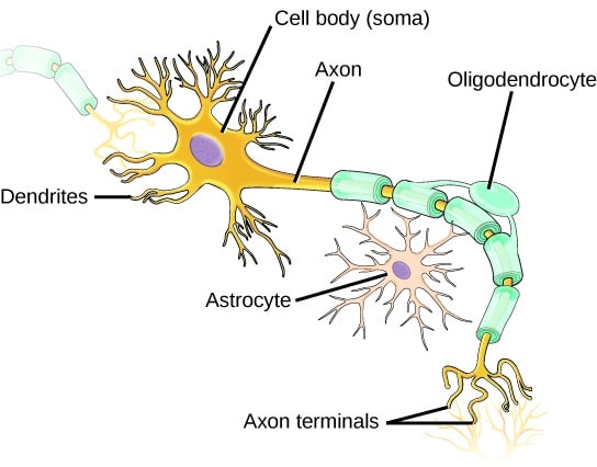
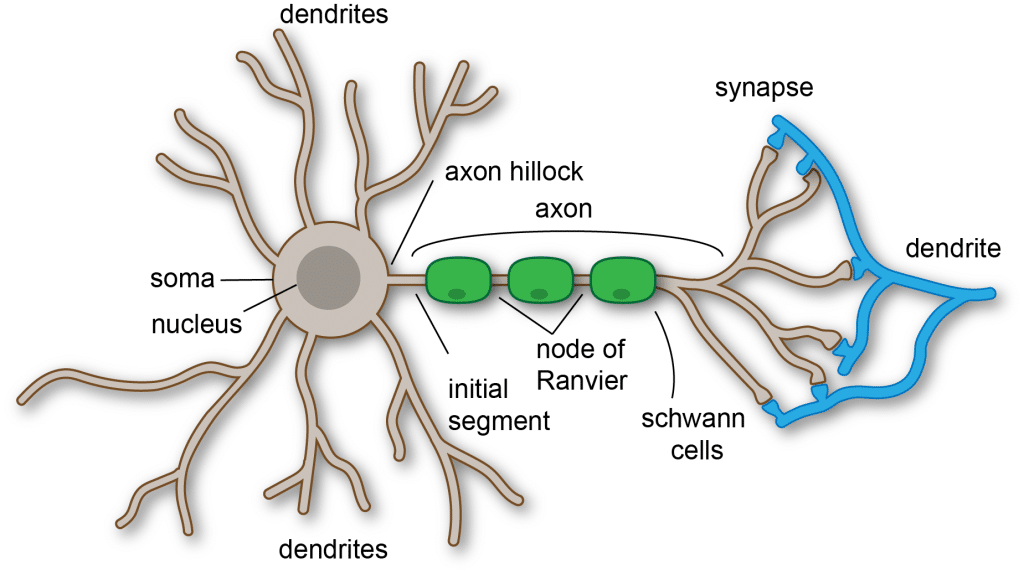
- Cell body or Soma – this contains the nucleus and the neurone’s intracellular organelles (such as the mitochondria and Golgi apparatus). It is the centre of neuronal metabolism. It also contains the Nissl Substance, which are granules containing rough endoplasmic reticulum and free ribosomes. This is the site of protein synthesis.
- Dendrites – these processes originate from the soma and extend outwards. They transmit signals received from other neurones to the soma.
- Axon – It arises from the soma, specifically from an area called the axon hillock, where action potentials are initiated. The action potentials are conducted along the axon to the axon terminal.
- Schwann cells – These insulate the axon with myelin sheath which facilitates the rapid transmission of action potentials along the axon.
- Axon terminal – Distally the axon branches, forming axon terminals. These make synaptic connections with other neurones. They contain various neurotransmitters which are released into the synapses to allow signal transmission from one neurone to the next.
Glial Cells
Astrocytes
Astrocytes are star-shaped glial cells within the brain and spinal cord, depending on the method used they make up between 20 and 40% of all glial cells. They have numerous functions, including:
- Metabolic support – The neurones have a constant requirement for nutrients such as glucose but they are unable to store or produce glycogen themselves. This is overcome by the fact that astrocytes store glycogen which can be broken down to glucose to provide fuel for neurones. Astrocytes can also store lactate which is useful as fuel during periods of high energy consumption or ischaemia.
- Regulation of extracellular ionic environment – High extracellular concentrations of ions such as potassium can result in spontaneous depolarisation of the neurone. Astrocytes prevent this by removing excess potassium ions from the extracellular space following neuronal activation.
- Neurotransmitter uptake – Astrocytes contain specific transporters for several neurotransmitters such as glutamate. A rapid removal of neurotransmitters from the extracellular space is required for the normal function of neurones.
- Modulation of synaptic transmission – In some regions of the brain, for example, the hippocampus, astrocytes release ATP in order to increase the production of adenosine, which in turn inhibits synaptic transmission. Hence, adenosine and other substances released by glial cells act as gliotransmitters modulating the synaptic activity.
- Promotion of myelination by oligodendrocytes.
Oligodendrocytes
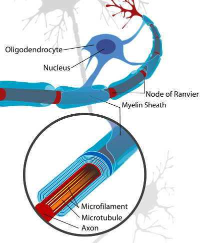
These cells are responsible for insulating the axons in the central nervous system. They carry out this function by producing a myelin sheath that wraps around a part of the axon.
A single oligodendrocyte has the capacity to myelinate up to 50 axonal segments. They are equivalent to the Schwann cells in the peripheral nervous system. Further information on the myelin sheath can be found here.

Fig 2 – Diagram showing the axon of a neurone in relation to the associated oligodendrocyte and myelin sheath
Microglia
Microglial cells make up between 10 and 15% of cells within the brain and are of a mesodermal origin, unlike the other glial cells which are of ectodermal origin.
These cells form the resident immune system of the brain. They are activated in response to tissue damage and have the capability to recognise foreign antigens and initiate phagocytosis to remove foreign material. If needed, microglia are also able to function as antigen-presenting cells.
Ependymal cells
The ependyma is the thin lining of the ventricular system of the brain and spinal cord. This lining is made up of ependymal cells, the basal membranes of which are attached to astrocytes. The main function of these cells is the production of cerebrospinal fluid (CSF) as a part of the choroid plexus.
Their apical surfaces are covered with cilia and microvilli, which allow for the circulation and absorption of CSF respectively.

Fig 3 – Diagram showing some of the glial cells in relation to a neuron
Clinical Relevance – Astrocytoma
Astrocytomas are intracranial tumours that are derived from astrocytes and may occur at any age, although they are more common in males.
They can be divided based on their grade which refers to the degree of their cellular differentiation:
- Low grade – Grade I and II, more common in children
- High grade – Grade III and IV, more common in adults
Low-grade astrocytomas are typically benign in nature and grow slowly. However, grade II tumours have the potential to become malignant. Grade I astrocytomas are often found within the cerebellum and therefore tend to present with symptoms relating to balance and coordination. Grade II tumours often present with seizures.
High-grade astrocytomas typically grow much more rapidly than low-grade ones and are generally malignant. Because of their invasive nature, they are often difficult to remove completely surgically and frequently recur after treatment.

